Research
Introduction:
This research asseses HRNet and ResNet architecture for their precision in localizing hand acupoints on 2D images, which is integral to automated acupunture therapy.
Objectives:
The primary objective was to advance the accuarcy of acupoint detection in traditional Korean medicine through the application of these advanced deep-learning models.
Background:
Acupoint localization in traditional Korean medicine is crucial for effective treatment, and the study aims to enhance this process usintg advanced deep-learning models.
Methods:
The study employs YOLOv3, YOLOF, and YOLOX-s for object detection within a top-down framework, comparing HRNet and ResNet architectures. These models were trained and tested using datasets annotated by technicians and their mean values, with performance evaluated based on Average Precision at two IoU thresholds.
Results:
HRNet consistently demonstrated lower mean distance errors across various acupoints compared to ResNet, particularly at a 256x256 pixel resolution. Notably, the HRNet-w48 model surpassed human annotators, including medical experts, in localization accuracy.
Conclusion:
HRNet's superior performance in acupoint localization suggests its potential to improve the precision and efficacy of acupuncture treatments. The study highlights the promising role of machine learning in enhancing traditional medical practices and underscores the importance of accurate acupoint localization in clinical acupuncture.
Real-time Location of Acupunture Points Based on Anatomical Landmarks and Pose Estimation Models
Precice identification of acupunture points (acupoints) is crucial for effective acupunture treatment, but manual localization by the unskilled can lack accuracy and consistency. This study aims to propose two computer vision approaches leveraging artificial intelligence (AI) for automated real-time localization and visualization of facial and hand acupoints. The firest approach employs a real-time landmark detection framework, detecting 38, acupoint locations on the face and hand by transforming anatomical landmark coordinates derived from image data. The second approach employs a state-of-the-art convolutional neural network optimized for pose estimation to detect five key arm and hand acupoints (LI11, LI10, TE5, TE3, LI4) in constrained medical imaging datasets. Valication against expert annotations demonstrated less than 5 mm mean localization errors for both approaches, indication high accuracy, These AI-driven techniques establish foundations for relable, automated acupoint recognition systems. Enabling self-localization of acupoints through imaging data focilitates self-training for individuals, provides assistive localization for practitioners, potentially improving the accuracy and accessibility of acupuncture treatments.
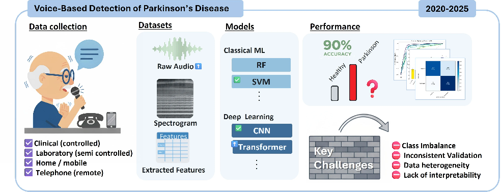
Speech impairments often emerge as one of the primary indicators of Parkinson’s disease (PD), albeit not readily apparent in its early stages. While previous studies focused predominantly on binary PD detection, this research explored the use of deep learning models to automatically classify sustained vowel recordings into healthy controls, mild PD, or severe PD based on motor symptom severity scores. Popular convolutional neural network (CNN) architectures, VGG and ResNet, as well as vision transformers, Swin, were fine-tuned on log mel spectrogram image representations of the segmented voice data. Furthermore, the research investigated the effects of audio segment lengths and specific vowel sounds on the performance of these models. The findings indicated that implementing longer segments yielded better performance. The models showed strong capability in distinguishing PD from healthy subjects, achieving over 95% precision. However, reliably discriminating between mild and severe PD cases remained challenging. The VGG16 achieved the best overall classification performance with 91.8% accuracy and the largest area under the ROC curve. Furthermore, focusing analysis on the vowel /u/ could further improve accuracy to 96%. Applying visualization techniques like Grad-CAM also highlighted how CNN models focused on localized spectrogram regions while transformers attended to more widespread patterns. Overall, this work showed the potential of deep learning for non-invasive screening and monitoring of PD progression from voice recordings, but larger multi-class labeled datasets are needed to further improve severity classification.
An analysis of scar tissue is necessary to understand the pathological tissue conditions during or after the wound healing process. Hematoxylin and eosin (HE) staining has conventionally been applied to understand the morphology of scar tissue. However, the scar lesions cannot be analyzed from a whole slide image. The current study aimed to develop a method for the rapid and automatic characterization of scar lesions in HE-stained scar tissues using a supervised and unsupervised learning algorithm. The supervised learning used a Mask region-based convolutional neural network (RCNN) to train a pattern from a data representation using MMDetection tools. The K-means algorithm characterized the HE-stained tissue and extracted the main features, such as the collagen density and directional variance of the collagen. The Mask RCNN model effectively predicted scar images using various backbone networks (e.g., ResNet50, ResNet101, ResNeSt50, and ResNeSt101) with high accuracy. The K-means clustering method successfully characterized the HE-stained tissue by separating the main features in terms of the collagen fiber and dermal mature components, namely, the glands, hair follicles, and nuclei. A quantitative analysis of the scar tissue in terms of the collagen density and directional variance of the collagen confirmed 50% differences between the normal and scar tissues. The proposed methods were utilized to characterize the pathological features of scar tissue for an objective histological analysis. The trained model is time efficient when used for detection in place of a manual analysis. Machine learning-assisted analysis is expected to aid in understanding scar conditions, and to help establish an optimal treatment plan.
Gating Mechanism of the Voltage-Gated Proton Channel Studied by Molecular Dynamics Simulations
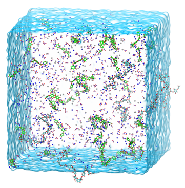
The voltage-gated proton channel Hv1 has important roles in proton extrusion, pH homeostasis, sperm motility, and cancer progression. The Hv1 channel has also been found to be highly expressed in cell lines and tissue samples from patients with breast cancer. A high-resolution closed state structure has been reported for the mouse Hv1 chimera channel (mHv1cc), solved by X-ray crystallography, but the open-state structure of Hv1 has not been solved. Since Hv1 is a promising drug target, various groups have proposed open conformations by molecular modeling and simulation studies. However, the gating mechanism and the open-state conformation under the membrane potential are still debate. Here, we present a molecular dynamics study considering membrane potential and pH conditions. The closed-state structure of mHv1cc was used to run molecular dynamics (MD) simulations with respect to electric field and pH conditions in order to investigate the mechanism of proton transfer. We observed a continuous hydrogen bond chain of water molecules called a water-wire to be formed through the channel pore in the channel opening, triggered by downward displacement of the S2 helix and upward movement of the S4 helix relative to other helices. Due to the movement of the S2 and S4 helices, the internal salt bridge network was rearranged, and the hydrophobic gating layers were destroyed. In line with previous experimental and simulation observations, our simulation results led us to propose a new gating mechanism for the Hv1 proton channel and may provide valuable information for novel drug discovery.
Functional stability of water wire-carbonyl interactions in an ion channel
Water wires are critical for the functioning of many membrane proteins, as in channels that conduct water, protons, and other ions. Here, in liquid crystalline lipid bilayers under symmetric environmental conditions, the selective hydrogen bonding interactions between eight waters comprising a water wire and a subset of 26 carbonyl oxygens lining the antiparallel dimeric gramicidin A channel are characterized by 17O NMR spectroscopy at 35.2 T (or 1,500 MHz for 1H) and computational studies. While backbone 15N spectra clearly indicate structural symmetry between the two subunits, single site 17O labels of the pore-lining carbonyls report two resonances, implying a break in dimer symmetry caused by the selective interactions with the water wire. The 17O shifts document selective water hydrogen bonding with carbonyl oxygens that are stable on the millisecond timescale. Such interactions are supported by density functional theory calculations on snapshots taken from molecular dynamics simulations. Water hydrogen bonding in the pore is restricted to just three simultaneous interactions, unlike bulk water environs. The stability of the water wire orientation and its electric dipole leads to opposite charge-dipole interactions for K+ ions bound at the two ends of the pore, thereby providing a simple explanation for an ∼20-fold difference in K+ affinity between two binding sites that are ∼24 ? apart. The 17O NMR spectroscopy reported here represents a breakthrough in high field NMR technology that will have applications throughout molecular biophysics, because of the acute sensitivity of the 17O nucleus to its chemical environment.
The inhibition of human angiotensin I converting enzyme (ACE) has been regarded as a promising approach for the treatment of hypertension. Despite research attempts over many years, our understanding the mechanisms of activation and inhibition of ACE is still far from complete. Here, we present results of all atom molecular dynamics simulations of ACE with and without ligands. Two types of inhibitors, competitive and mixed non-competitive, were used to model the ligand bound forms. In the absence of a ligand the simulation showed spontaneous large hinge-bending motions of multiple conversions between the closed and open states of ACE, while the ligand bound forms were stable in the closed state. Our simulation results imply that the equilibrium between pre-existing backbone conformations shifts in the presence of a ligand. The hinge-bending motion of ACE is considered as an essential to the enzyme function. A mechanistic model of activation and the inhibition may provide valuable information for novel inhibitors of ACE.
In cellular environments, proteins not only interact with their specific partners but also encounter a high concentration of bystander macromolecules, or crowders. Nonspecific interactions with macromolecular crowders modulate the activities of proteins, but our knowledge about the rules of nonspecific interactions is still very limited. In previous work, we presented experimental evidence that macromolecular crowders acted competitively in inhibiting the binding of maltose binding protein (MBP) with its ligand maltose. Competition between a ligand and an inhibitor may result from binding to either the same site or different conformations of the protein. Maltose binds to the cleft between two lobes of MBP, and in a series of mutants, the affinities increased with an increase in the extent of lobe closure. Here we investigated whether macromolecular crowders also have a conformational or site preference when binding to MBP. The affinities of a polymer crowder, Ficoll70, measured by monitoring tryptophan fluorescence were 3?6-fold higher for closure mutants than for wild-type MBP. Competition between the ligand and crowder, as indicated by fitting of titration data and directly by nuclear magnetic resonance spectroscopy, and their similar preferences for closed MBP conformations further suggest the scenario in which the crowder, like maltose, preferentially binds to the interlobe cleft of MBP. Similar observations were made for bovine serum albumin as a protein crowder. Conformational and site preferences in MBP?crowder binding allude to the paradigm that nonspecific interactions can possess hallmarks of molecular recognition, which may be essential for intracellular organizations including colocalization of proteins and liquid?liquid phase separation.
In this work, a target-based drug screening method is proposed exploiting the synergy effect of ligandbased and structure-based computer-assisted drug design. The new method provides great flexibility in drug design and drug candidates with considerably lower risk in an efficient manner. As a model system, 45 sulphonamides (33 training, 12 testing ligands) in complex with carbonic anhydrase IX were used for development of quantitative structure-activity-lipophilicity (property)-relationships (QSPRs). For each ligand, nearly 5,000 molecular descriptors were calculated, while lipophilicity (logkw) and inhibitory activity (logKi) were used as drug properties. Genetic algorithm-partial least squares (GA-PLS) provided a QSPR model with high prediction capability employing only seven molecular descriptors. As a proof-of-concept, optimal drug structure was obtained by inverting the model with respect to reference drug properties. 3509 ligands were ranked accordingly. Top 10 ligands were further validated through molecular docking. Large-scale MD simulations were performed to test the stability of structures of selected ligands obtained through docking complemented with biophysical experiments.
Silver-Lactoferrin Nanocomplexes as a Potent Antimicrobial Agent
The process of silver immobilization onto and/or into bovine lactoferrin (LTF), the physicochemical properties of bovine lactoferrin and obtained silver-lactoferrin complexes, as well as antibacterial activity of silver-lactoferrin complexes were investigated in this work. Kinetic study of the silver immobilization into lactoferrin was carried out using batch sorption techniques. Spectrometric (MALDI-TOF/TOF-MS, ICP-MS), spectroscopic (FTIR, SERS), electron microscopic (TEM) and electrophoretic (I-DE) techniques, as well as zeta potential measurements, were applied for characterization of LTF and binding nature of silver in Ag-LTF complexes. On the basis of the results of the kinetics study, it was established that the silver binding to LTF is a heterogeneous process involving two main stages: (i) internal diffusion and sorption onto external surface of lactoferrin globules; and (ii) internal diffusion and binding into lactoferrin globule structure. Spectroscopic techniques combined with TEM analysis confirmed the binding process. Molecular dynamics (MD) analysis was carried out in order to simulate the mechanism of the binding process, and locate potential binding sites, as well as complement the experimental findings. Quantum mechanics (QM) simulations were performed utilizing density functional theory (DFT) in order to support the reduction mechanism of silver ions to elemental silver. Antimicrobial activity of synthesized lactoferrin complexes against selected clinical bacteria was confirmed using flow cytometry and antibiograms.
High-speed signal processing is essential for real-time displays in medical imaging applications. Photoacoustic tomography provides structural, functional, and molecular imaging with high resolution in a noninvasive way. Especially, three-dimensional image reconstruction, functional imaging, and real-time display require fast signal processing. Here, we provide a high-speed signal processing method using a graphic processing unit (GPU) to reconstruct ultrasound or photoacoustic B-mode images for real-time displays. The signal processing speed wat improved by parallel processing of the beam formation and the envelop detection required for image reconstruction using a massive number of GPU cores. The time suing a GPU was 2.778 ms, on average, to process a single-frame B-mode image with 128 x 3200 pixels while it was about 2.165 seconds using a central processing unit (CPU). The processing time suing a GPU was short enough the reconstruct three-dimensional images for real-time displays.
Insight into the mechanism of the influenza A proton channel from a structure in a lipid bilayer (PDB deposit: 2L0J).
The M2 protein from the influenza A virus, an acid-activated proton-selective channel, has been the subject of numerous conductance, structural, and computational studies. However, little is known at the atomic level about the heart of the functional mechanism for this tetrameric protein, a His37-Trp41 cluster. We report the structure of the M2 conductance domain (residues 22 to 62) in a lipid bilayer, which displays the defining features of the native protein that have not been attainable from structures solubilized by detergents. We propose that the tetrameric His37-Trp41 cluster guides protons through the channel by forming and breaking hydrogen bonds between adjacent pairs of histidines and through specific interactions of the histidines with the tryptophan gate. This mechanism explains the main observations on M2 proton conductance.
Conformational heterogeneity of the M2 proton channel and a structural model for channel activation.
The M2 protein of influenza virus A is a proton-selective ion channel activated by pH. Structure determination by solid-state and solution NMR and X-ray crystallography has contributed significantly to our understanding, but channel activation may involve conformations not captured by these studies. Indeed, solidstate NMR data demonstrate that the M2 protein possesses significant conformational heterogeneity. Here, we report molecular dynamics (MD) simulations of the M2 transmembrane domain (TMD) in the absence and presence of the antiviral drug amantadine. The ensembles of MD conformations for both apo and bound forms reproduced the NMR data well. The TMD helix was found to kink around Gly-34, where water molecules penetrated deeply into the backbone. The amantadine-bound form exhibited a single peak ~10° in the distribution of helix-kink angle, but the apo form exhibited 2 peaks, ~0° and 40°. Conformations of the apo form with small and large kink angles had narrow and wide pores, respectively, around the primary gate formed by His-37 and Trp-41. We propose a structural model for channel activation, in which the small-kink conformations dominate before proton uptake by His-37 from the exterior, and proton uptake makes the large-kink conformations more favorable, thereby priming His-37 for proton release to the interior.
A wealth of experimental data has verified the applicability of the Gouy-Chapman (GC) theory to charged lipid membranes. Surprisingly, a validation of GC by molecular dynamics (MD) simulations has been elusive. Here, we report a test of GC against extensive MD simulations of an anionic lipid bilayer solvated by water at different concentrations of NaCl or KCl. We demonstrate that the ion distributions from the simulations agree remarkably well with GC predictions when information on the adsorption of counterions to the bilayer is incorporated. A wealth of experimental data has verified the applicability of the Gouy-Chapman (GC) theory to charged lipid membranes. Surprisingly, a validation of GC by molecular dynamics (MD) simulations has been elusive. Here, we report a test of GC against extensive MD simulations of an anionic lipid bilayer solvated by water at different concentrations of NaCl or KCl. We demonstrate that the ion distributions from the simulations agree remarkably well with GC predictions when information on the adsorption of counterions to the bilayer is incorporated.
Nicotinic AChRs (nAChRs) represent a paradigm for ligand-gated ion channels. Despite intensive studies over many years, our understanding of the mechanisms of activation and inhibition for nAChRs is still incomplete. Here, we present molecular dynamics (MD) simulations of the alpha7 nAChR ligand-binding domain, both in apo form and in alpha-Cobratoxin-bound form, starting from the respective homology models built on crystal structures of the acetylcholine-binding protein. The toxin-bound form was relatively stable, and its structure was validated by calculating mutational effects on the toxin-binding affinity. However, in the apo form, one subunit spontaneously moved away from the conformation of the other four subunits. This motion resembles what has been proposed for leading to channel opening. At the top, the C loop and the adjacent beta7-beta8 loop swing downward and inward, whereas at the bottom, the F loop and the C terminus of beta10 swing in the opposite direction. These swings appear to tilt the whole subunit clockwise. The resulting changes in solvent accessibility show strong correlation with experimental results by the substituted cysteine accessibility method upon addition of acetylcholine. Our MD simulation results suggest a mechanistic model in which the apo form, although predominantly sampling the ‘‘closed’’ state, can make excursions into the ‘‘open’’ state. The open state has high affinity for agonists, leading to channel activation, whereas the closed state upon distortion has high affinity for antagonists, leading to inhibition.